Clinical History
A 5-year-old female presented with a complaint of urinary retention.
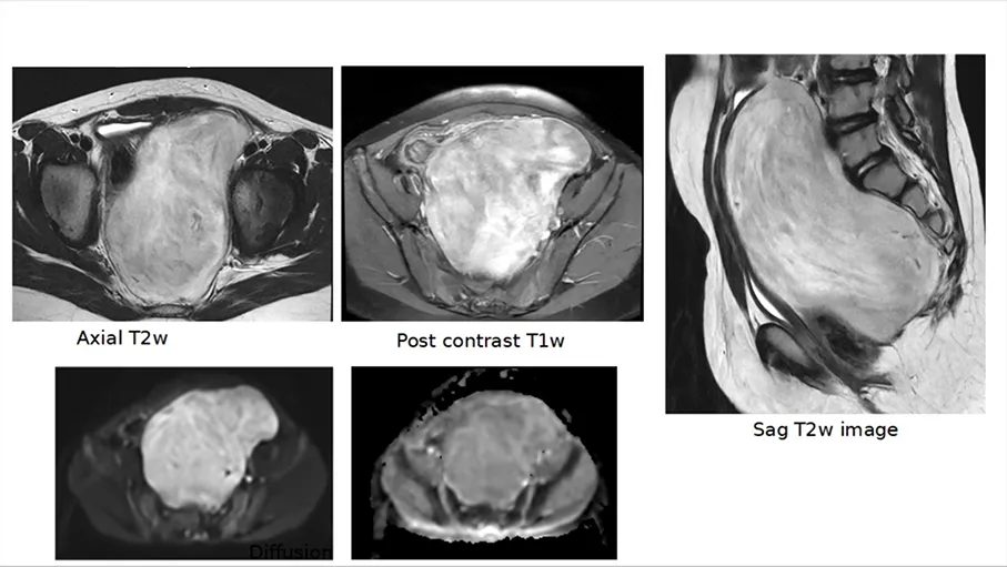
Ultrasound examination revealed a large, well-defined heterogeneous pelvic lesion measuring 11.7 × 9.8 × 6.6 cm, demonstrating internal vascularity. The lesion was noted to be displacing both ovaries and the urinary bladder superiorly.
Serum Alpha-Fetoprotein (AFP) and Beta Human Chorionic Gonadotropin (β-HCG) levels were within normal limits.
Diagnosis Answer
Findings: Location of the mass was presacral. Urinary bladder was displaced anteriorly. Rectum displaced anteriorly and to the right ( arrow). Bone was not involved. No extension to the sacral foramina seen. Biopsy was performed- s/o Ganglioneuroma Common Differential diagnosis of a presacral mass in this age are: Congenital- cystic hamartoma, duplication cyst, dermoid / germ cell tumour or anterior meningocele Osseous- Ewing’s sarcoma, ABC Mesenchymal- sarcoma Neurogenic masses- neuroblastoma, ganglioneuroma, ganglioneuroblastoma, nerve sheath tumour Others- DSRCT, lymphoma.