Indian Society of Paediatric Radiology

UPCOMING EC MEETING
WHO WE ARE

The Indian Society of Paediatric Radiology (ISPR) was constituted to encourage and advance the knowledge and study of Paediatric Radiology in India since the vast knowledge of medical science needs to look at children and their medical problems in a different perspective. Diseases of children are completely different from adults including the anatomical make up of children differs from that of adults. India being the second most populous country in the world has a considerable paediatric age group population.

The society was formed so that dedicated paediatric radiologists can help, teach and promulgate the knowledge of this discipline to young and upcoming radiologists and help bridge the gap of such radiologists in our institutions which have a lack of specialized radioliogists. It is a non-profitable organization for the promotion of paediatric radiology in India and abroad to assist Government and Non-governmental agencies in all matters pertaining to paediatric radiology & medical diagnostic imaging, to encourage scientific work as well as to promote interaction among members of ISPR and allied specialists.

We hold one Mid-term National CME and one National Conference a year.
We publish an E-Newsletter twice a year for all our members.
We interact with other societies in India & in the world such as WFPI (World Federation of Paediatric Imaging) and AOSPR (Asian & Oceanic Society for Paediatric Radiology).
We have a dedicated team of office bearers with ISPR who help to carry the flag of ISPR forward.
QUIZ
MAY 2024 QUIZ
MAY QUIZ ANSWER PAGE -> LIST OF PARTICIPANTS ->
JUNE 2024 QUIZ
NEW QUIZ-->

Quiz Answer May 2024

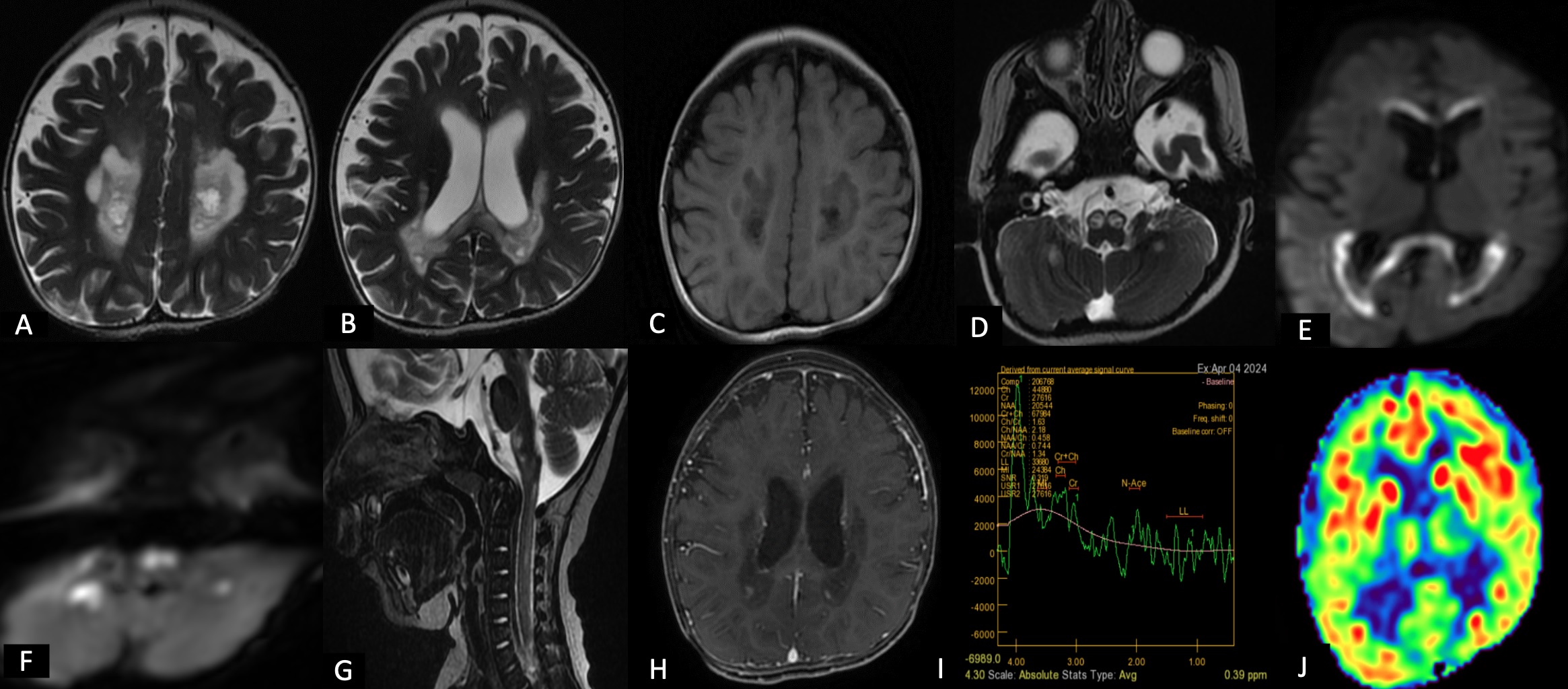
Diagnosis

  • Succinate Dehydrogenase Related
  • Leukoencephalopathy (Complex ii/Succinate-ubiquinone Oxidoreductase Deficiency)

qaMay2401 May 2024

IMAGING FEATURES

  • Involvement of specific thalamic nuclei, the cerebral hemispheric WM abnormalities with sparing of the U fibers, Corpus callosum involvement with sparing of the outer blades, and the cervical spinal cord abnormalities - strongly suggestive of SDH deficiency related leukoencephalopathy.
  • Involvement of the corticospinal tracts - posterior limb of the internal capsule, the pons, and the pyramids of the medulla is also seen.
  • The corticopontocerebellar tracts are also frequently affected, with signal abnormalities in the corticopontine and transverse pontine fibers and middle cerebellar peduncles.
  • MRS (short echo time) may show a sizeable metabolic peak at 2.4ppm representing succinate. Also, a macromolecular peak centered at 1.3ppm consistent with lactate can be found.
  • Acute stage - abnormal WM looks swollen, especially the corpus callosum with lesions showing restricted diffusion.
  • Intermediate stage - WM rarefaction and small foci of contrast enhancement may be observed. No restricted diffusion foci are noted.
  • Late stage- atrophy and collapse of the affected WM are seen, with cysts.

DIFFERENTIAL DIAGNOSIS

  1. Leukoencephalopathy with Brainstem and Spinal Cord Involvement and High Lactate (LBSL)
    - Compound heterozygous mutations in the gene DARS2 which encodes for mitochondrial aspartyl-tRNA synthetase
    - Imaging findings: Symmetric involvement of WM, sparing subcortical WM
    - Lesions in the mesencephalic trigeminal tract, inferior cerebellar peduncles, and splenium of the corpus callosum
  2. Vanishing white matter disease (VWM): Bilateral symmetrical involvement of periventricular white matter with
    involvement of subcortical U fibers and is replaced with fluid attenuating areas evidence of any hyperintensity in brain
    stem and cerebellum. Normal lactate levels.
  3. Prenyldiphosphate synthase subset 1(PDSS 1) deficiency : Bilateral periventricular white matter hyperintensity with
    cystic change with involvement of corpus callosum and porencephalic cyst formation

MANAGEMENT

  1. No standard treatment algorithm currently exists.
  2. Riboflavin has been shown to alleviate some of the symptoms and delay disease progression.
  3. Oral coenzyme Q10 as treatment.

REFERENCES

  1. Augusto C, Fabricio Guimaraes Goncalves, Grieb D, Leandro Tavares Lucato, Goldstein AC, Giulio Zuccoli. Neuroimaging of Mitochondrial Cytopathies. Topics in magnetic resonance imaging. 2018 Aug 1;27(4):219-40
  2. Alston CL, Davison JE, Meloni F, van der Westhuizen FH, He L, Hornig-Do HT, et al. Recessive germline SDHA and SDHB mutations causing leukodystrophy and isolated mitochondrial complex II deficiency. Journal of Medical Genetics. 2012 Sep;49(9):569-77.
AFFILIATES